
Novel 3D imaging for chest wall anomalies in Calgary
| Novel 3D imaging for chest wall anomalies: The early Calgary experience, Jennifer YK Lam, Janet Ronsky, Marc Schneider, Mary Brindle, Steven Lopushinsky, Nancy Schneider, Jacob Reichbart. Canadian Association of Paediatric Surgeons CAPS Annual Meeting, Banff AB October 2017. Video by Christina Ryan Productions. Youtube Oct 06, 2017 |
| Purpose |
Clinical measurements of pectus carinatum deformity vary by clinician, often performed with tape measure or calipers. We propose the use of three dimensional body scans to quantify chest dimensions and severity of pectus deformities.
| Methods |
Patients were recruited for study at time of brace fitting for pectus carinatum (ethics approved). Patients were measured by an experienced clinician with calipers to identify the anterior-posterior and transverse dimensions of the chest. A three dimensional body scan was performed, converted to an axial image and deformity dimensions were obtained for comparison (Figure 1). Pearson correlation coefficients were calculated.
| Results |
Between October 2016 and February 2017, 13 male patients were enrolled at initiation of pectus carinatum treatment (median age 14, range 12-17). The calculated Pearson correlation coefficients for anterior-posterior and transverse chest dimensions were 0.7 and 0.5, respectively.
| Conclusion |
There is positive correlation between three dimensional body scan and clinical measurements. Three dimensional scanning is easily implemented in the clinic setting and may be a more objective measure of deformity severity and treatment success over time. Future study is required to better characterize individualized pectus carinatum therapy as well as a potential role in reducing ionizing radiation in the work up of pectus excavatum.
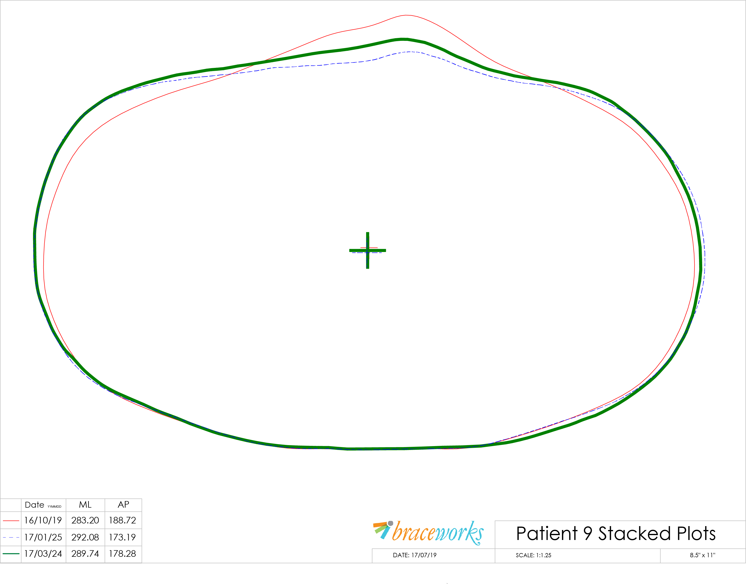
| Result of wearing the Braceworks® Pectus Brace over time. Stacked plot shows reshaping of the chest wall early in treatment — 3 transverse sections taken at a given T-level. |
| References |
Three dimensional body scans for an objective measurement of pectus carinatum deformities, Jennifer YK Lam, Janet Ronsky, Marc Schneider, Mary Brindle, Steven Lopushinsky. Abstract 45, Canadian Association Of Paediatric Surgeons CAPS 49th Annual Meeting Banff, Alberta Canada, October 5-7, 2017
Dynamic bracing of pectus carinatum: a quantitative analysis, Tomasz Bugajski, Kartikeya Murari, Steven Lopushinsky, Marc Schneider, Jacob Reichbart, Janet Ronsky. Abstract 19, Canadian Association Of Paediatric Surgeons CAPS 49th Annual Meeting Banff, Alberta Canada, October 5-7, 2017
Reliability of a Three-Dimensional Scanning Technique and Metrics Quantifying Pectus Deformities, Tomasz Bugajski, Bahareh Vafadar, Emma Gray, Kopal Garg, Marc Schneider, Alberto Nettel-Aguirre, Mary Brindle, Jennifer Lam, Steven Lopushinsky, Janet Ronsky. Schulich School of Engineering, University of Calgary. Braceworks® Custom Orthotics Ltd, Calgary. Alberta Children’s Hospital, Calgary AB Canada. 2017
| Further reading |
New Methods for Imaging Evaluation of Chest Wall Deformities, Ana Lain, Laura Garcia, Carlos Gine, Olivier Tiffet, and Manuel Lopez. Front. Pediatr. 04 December 2017. doi: 10.3389/fped.2017.00257
Evaluation of the treatment of pectus carinatum with compressive orthotic bracing using three dimensional body scans, Wong KE, Gorton GE 3rd, Tashjian DB, Tirabassi MV, Moriarty KP. J Pediatr Surg. 2014 Jun;49(6):924-7. doi: 10.1016/j.jpedsurg.2014.01.024. Epub 2014 Feb 3.
Does an external chest wall measurement correlate with a CT-based measurement in patients with chest wall deformities? Ewert F, Syed J, Wagner S, Besendoerfer M, Carbon RT, Schulz-Drost S. J Pediatr Surg. 2017 Oct;52(10):1583-1590. doi: 10.1016/j.jpedsurg.2017.04.011. Epub 2017 Apr 27.
A Simplified Method for Three-Dimensional Optical Imaging and Measurement of Patients with Chest Wall Deformities, Szafer D, Taylor JS, Pei A, de Ruijter V, Hosseini H, Chao S, Wall J. J Laparoendosc Adv Surg Tech A. 2019 Feb;29(2):267-271. doi: 10.1089/lap.2018.0191. Epub 2018 Sep 12.