
A new wearable brain scanner
A helmet records a wearer’s brain activity using magnetoencephalography (MEG) while they move around.
In a design that looks straight out of an old future-tech horror film, researchers in the U.K. have built a wearable, portable brain scanner that can record neural activity while the user is moving.

University of Nottingham photo
By Emily Waltz, IEEE Spectrum 21 March 2018
The device, described today in Nature, could enable scientists to study brain function in ways that aren’t possible with stationary brain scanners, like that of functional magnetic resonance imaging, or fMRI.
“It’s a big step forward,” says Peter Schwindt, a physicist at Sandia National Laboratories in Albuquerque NM, who was not involved in the project. The technology “opens up new applications” for this type of brain scanning, he says.
That assumes people can get over the unfortunate look of the device, a wired-up head cast that falls somewhere between The Phantom of the Opera and Predator.
The device employs magnetoencephalography, or MEG, which measures magnetic fields present at the scalp. These fields are generated by the brain’s natural electrical currents, and, with mathematical analysis, can be used to create a 3D map of brain function with millisecond resolution.
Conventional MEG devices—cumbersome machines the size of a manatee—require the user to remain motionless while undergoing a scan, similar to the requirements of an fMRI. That severely limits the kinds of research that can be conducted. It also makes it difficult to study children.
In today’s report, researchers at the University of Nottingham and University College London, in the U.K., shrunk MEG to the size of gladiator helmet. The system would enable researchers to image people who find it hard to keep still, such as babies, children, and people with movement disorders.
The portable system would also allow scientists to conduct entirely new kinds of studies. “You can look at aspects of brain function involving spatial navigation, which is hard to do with a subject who is stationary,” says Richard Bowtell, a professor of physics at the University of Nottingham, who co-authored the report. “You can also look at more natural interactions between people when they are free to move.”
In the team’s design, the sensors are fixed relative to the person’s brain, rather than in a stationary machine. They achieved this by integrating miniaturized quantum sensors into a head cast, and pairing it with a system for canceling out background magnetic fields.

These helmets contain small sensors called magnetometers that detect magnetic fields to allow researchers to map a wearer’s brain activity. University of Nottingham
The system is custom made for each user. A head cast is 3D printed to fit snugly over the scalp and face. Miniaturized quantum sensors called optically pumped magnetometers (OPMs) lock in place above the target area of the brain, where they will sense the brain’s magnetic fields. (The sensors are commercially available through QuSpin in Louisville, Colorado).
To cancel out the Earth’s magnetic fields, which would interfere with the scan, the researchers constructed a set of bi-planar electromagnetic coils. These coils generate fields equal and opposite to the Earth’s field, thereby canceling it out. The coils are placed in a structure that sits near the user, creating a small, magnetically shielded space in which the user can move during the scan. The experiments take place in a magnetically shielded room which cancels additional fields.
Bowtell and his colleagues tested the system against a conventional MEG machine by recording subjects’ brain activity while they performed a finger lifting task. The wearable system performed on par with the conventional machine, according to today’s report.
The team then recorded the subjects’ brain activity while they performed different tasks that involve head movement, such as bouncing a ball on a paddle or drinking from a mug. “I was impressed by what they could do with measuring the brain response while playing this ball game,” says Schwindt at Sandia.
| Designing a new brain scanner (MEG). Scientists at the University of Nottingham are working with University College London (UCL) on a five-year project which has the potential to revolutionise the world of human brain imaging. Magnetoencephalography (MEG) is a technique for mapping brain activity – it measures the magnetic fields generated by electrical currents that occur naturally in the brain.
A £1.6m Collaborative Award in Science from Wellcome is funding the construction of a new type of MEG scanner which, if successful, could quadruple the sensitivity of current devices. The project is a collaboration between the University of Nottingham and University College London. Find out more about our collaborative awards in science. Wellcome Trust. Uploaded Youtube on Mar 13, 2018 |
The big limitation of the prototype is that users can’t move their heads outside of the shielded space: an invisible box 20 to 40 cm per side, or about the size of an old, venerable Macintosh SE. “Subjects are constrained by this 40 cm volume, so obviously they’re not getting up and walking around,” says Schwindt. “There’s significant development that needs to happen to move towards allowing full natural movement.”
Bowtell says his team is working on that. In the next iteration, the group aims to integrate the background-canceling coils into the walls of the room, allowing the subject to walk around.
Several groups, including Schwindt’s, have been developing quantum sensors, and specifically OPMs, for use in MEG imaging. OPMs improve MEG imaging because they don’t have to be cryogenically cooled, like the superconducting technology in conventional MEG scanner. That allows the OPM sensors to be worn snugly on the head, improving the quality of the data recorded.
Despite the improvements in OPM sensors, subjects must remain still during scans. “Most of us have taken the approach thus far to keep our sensors stationary,” Schwindt says.
The U.K. team is likely the first to employ OPM technology in a way that allows subjects to move, he says.
The idea of making brain recording and imaging devices more portable is not new, of course. Researchers have successfully built wearable EEG, or electroencephalography, and even used such devices to record the brain’s electrical activity during a bungee jumping experiment. EEG measures the voltages at the scalp, which reflects the voltages in the brain. But it’s hard to use EEG to pinpoint the location of the activity in the brain—something that MEG can do.
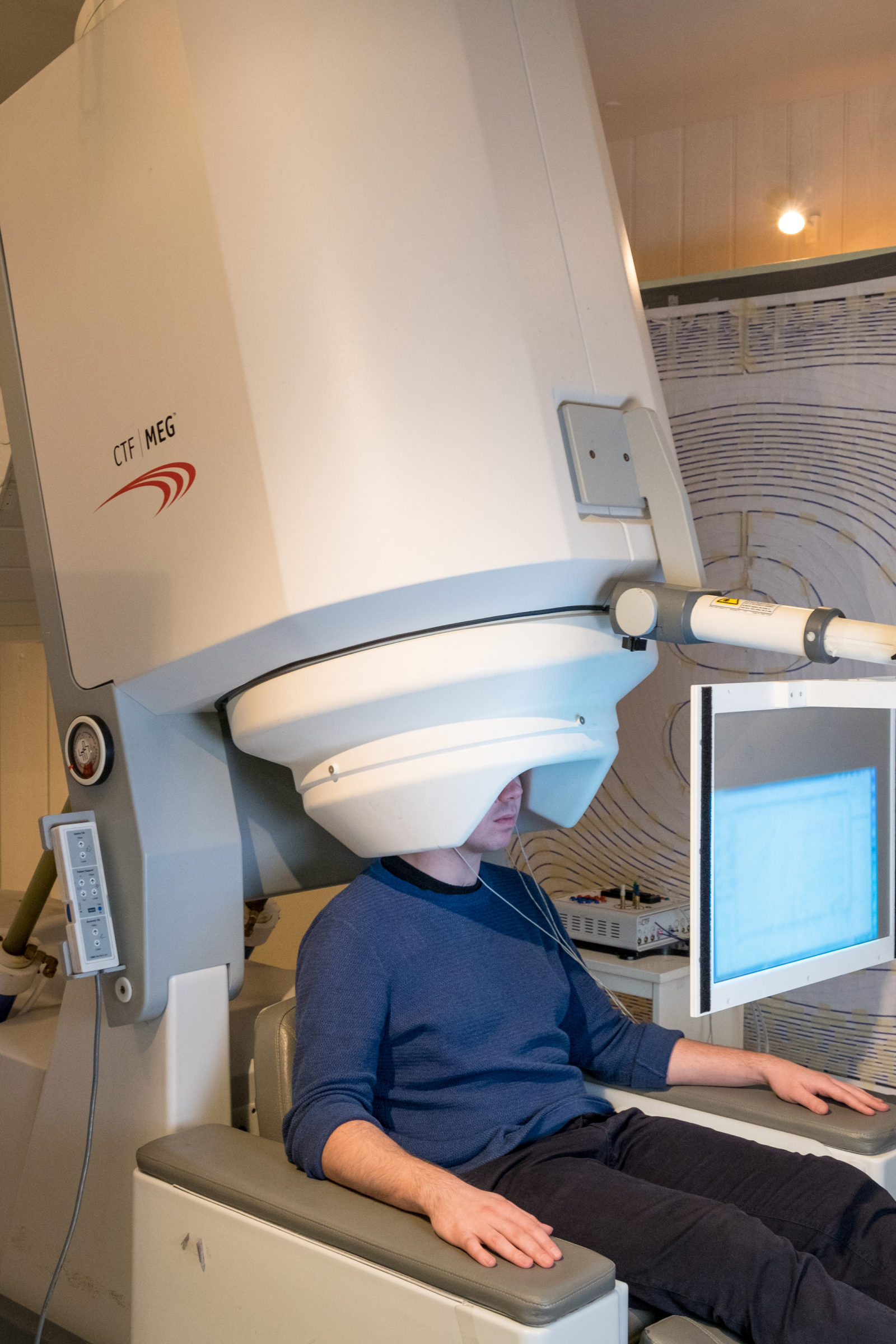
Current MEG scanners are large and weigh around half a tonne. This is because the sensors used to measure the brain’s magnetic field need to be kept very cold (-269°C) which requires bulky cooling technology. With current scanners, the patient must remain very still whilst being scanned, as even a 5-mm movement can make the images unusable. This means it is often difficult to scan people who find it hard to remain still such as young children, or patients with movement disorders. It also poses problems when one might need a patient to remain still for a long time in order to capture a rarely occurring event in the brain, such as an epileptic seizure. Wellcome Trust.
Researchers have also developed wearable brain scanners using fNIRS, or functional near-infrared spectroscopy. One group used the technique to create a brain-computer interface system. In fNIRS, changes in blood oxygenation are measured using light as an indirect indicator of neural activity. But like EEG, it doesn’t easily pinpoint the location of the brain activity, says Bowtell.
Wearable MEG could provide that specificity in a portable scenario. “It will be interesting to see how far the technology can be pushed, in terms of how much movement” can be allowed during scanning, says Schwindt.
And if the head cast ends up not working for the research world, maybe someone in Hollywood could use a new prop.
Source IEEE Spectrum
| References |
Moving magnetoencephalography towards real-world applications with a wearable system, Elena Boto, Niall Holmes, James Leggett, Gillian Roberts, Vishal Shah, Sofie S. Meyer, Leonardo Duque Muñoz, Karen J. Mullinger, Tim M. Tierney, Sven Bestmann, Gareth R. Barnes, Richard Bowtell & Matthew J. Brookes. Nature 21 March 2018 doi:10.1038/nature26147
A new generation of magnetoencephalography: Room temperature measurements using optically-pumped magnetometers, Boto E, Meyer SS, Shah V, Alem O, Knappe S, Kruger P, Fromhold TM, Lim M, Glover PM, Morris PG, Bowtell R, Barnes GR, Brookes MJ. Neuroimage. 2017 Apr 1;149:404-414. doi: 10.1016/j.neuroimage.2017.01.034. Epub 2017 Jan 25.
Also see
New brain scanner allows patients to move freely for the first time Wellcome Trust
In-Ear EEG Makes Unobtrusive Brain-Hacking Gadgets a Real Possibility IEEE Spectrum
Wireless Earbuds Will Record Your EEG, Send Brainwave Data to Your Phone IEEE Spectrum
Wearable Brain Scanner Tells Your Computer When You’re Overwhelmed IEEE Spectrum
Measuring Free Will of Bungee Jumpers IEEE Spectrum
This wearable brain scanner could transform our understanding of how neurons ‘talk’ Science AAAS
Helmet-shaped brain scanner allows wearers to move around The Guardian
Worn like a helmet, a new brain scanner aims to make it easier to treat kids with epilepsy STAT
Quantum sensors herald new generation of wearable brain imaging systems University of Nottingham